Summarize content with
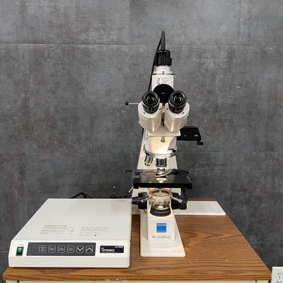
A Lab microscope condenser is a small but important part of a microscope. It sits right under the stage, and its job is to shine light onto the specimen you're looking at. This light helps make the image brighter and clearer.
You can think of the condenser like a spotlight. It takes the light from the microscope’s bulb, focuses it into a cone shape, and aims it right at the sample. This helps you see tiny things like cells or bacteria much more clearly.
Many condensers have a little part called an iris diaphragm. This part lets you adjust how much light goes through. By changing it, you can make the image lighter or darker, or adjust how much detail you can see. For example, if you're looking at something clear, changing the diaphragm can help you see it better.
There are also different kinds of condensers. Some are simple, like the Abbe condenser, which is common in school microscopes. Others are more advanced and used in science labs. These special ones can help reduce blurriness or show tiny details better.
So, even though the condenser is small, it plays a big role in helping you get a good look at your sample. Without it, the image might be too dark or blurry to study properly.
Why Does the Condenser Matters in Microscopy?
When you're using a microscope, seeing things clearly depends on more than just the lenses. One part that's really important but often overlooked is the condenser. It's the piece right under the stage that helps light shine onto the sample in just the right way.
The condenser takes light from the bulb and focuses it into a cone-shaped beam that goes through the specimen. This focused light helps make the image bright, clear, and full of detail. Without it, the picture might look too dim or blurry, even with a powerful lens.
Most condensers have a small part called an iris diaphragm. It works like a camera lens, letting you change how wide or narrow the light beam is. Opening it wide gives you more detail but less contrast. Closing it helps boost contrast, which is great when looking at clear samples like live cells.
The numerical aperture (NA) of the condenser is also very important. It tells you how much light the condenser can focus. To get the best image, the condenser’s NA should match or be slightly higher than the lens you're using. If it's too low, you won’t see the tiny details clearly.
Another thing the condenser helps with is Köhler illumination. This is a method scientists use to make the light even and smooth across what you're looking at. It helps get rid of shadows and weird bright spots, which is important when taking pictures or doing serious lab work.
The condenser is also needed for some special types of microscopy:
- Phase Contrast helps you see clear objects by turning invisible light changes into visible contrast. It needs a special ring in the condenser.
- Darkfield makes the background black and lights up the object only. It uses a condenser that blocks direct light.
- DIC (Differential Interference Contrast) makes samples look 3D by using special light tricks that need exact condenser alignment.
There are different types of condensers, too. A basic Abbe condenser is common in schools, but for clearer and more accurate images, especially with color, scientists use better versions like achromatic or aplanatic condensers. These reduce color errors and image blur.
Also, the condenser has to be lined up correctly. If it’s too low, off-center, or not adjusted properly, your image can look uneven, have strange halos, or lose sharpness. Some high-end microscopes even have oil-immersion condensers that work with special lenses to give maximum detail.
How a Microscope Condenser Works?
A microscope condenser might look like a small part, but it plays a big role in helping you see clear and detailed images. It’s the part that manages light collecting it, focusing it, and shining it just right onto your sample. Let’s walk through how it works in simple steps.
Step 1: Light Comes from the Bulb
Every microscope has a light source, like a small bulb or LED. This light spreads out in all directions, so it needs help to become useful for seeing tiny things.
Step 2: The Collector Lens Gathers the Light
A lens gathers this scattered light and sends it toward the next part of the microscope, the condenser’s diaphragm. This helps start shaping the light into a more focused beam.
Step 3: The Iris Diaphragm Adjusts the Light
Right before the light goes through the condenser lenses, it passes through a small opening called the iris diaphragm. This opening can be made wider or narrower:
- A wide opening gives you more detail but less contrast.
- A narrow one gives more contrast but might hide some small details.
Step 4: Condenser Lenses Focus the Light into a Cone
The condenser lenses take the light and bend it into a cone shape that points directly at your sample. The angle of this cone matters; it should match the power of the lens you’re using to get the best result.
Step 5: The Field Diaphragm Controls What Gets Lit
The condenser also helps decide how much of your sample area gets lit. There’s another part called the field diaphragm that you can open or close to control the size of the lit area. This helps reduce glare and makes the image clearer.
Step 6: Aligning the Condenser
To get the best view, the condenser must be in the right spot:
- Raise or lower it until the light hits the sample just right.
- Use small screws to center the light path.
- Adjust things so the field of view is evenly lit and not too bright or too dark.
This is part of what’s called Köhler illumination, which gives smooth, even lighting and helps you avoid bright spots or shadows.
Step 7: Light Goes Through the Specimen
The focused cone of light now passes through your sample. Stained parts of the sample may block some light, and clear parts might bend it. That’s how we start to see the shapes and details.
Step 8: The Objective Lens Creates the Image
After passing through the sample, the light moves up into the objective lens, which magnifies the image so you can see it through the eyepiece or camera.
Step 9: Adjust for Different Lenses
If you switch to a new objective lens (like from 10x to 100x), you also need to:
- Move the condenser up or down
- Adjust the iris diaphragm so the light fits that lens’s needs
If the light cone is too narrow, the image won’t be sharp. If it’s too wide, the contrast drops.
Step 10: Special Techniques Need the Condenser Too
The condenser helps with more than just basic viewing. Some special types of microscopy depend on it:
- Phase Contrast: Helps see clear samples using a special ring in the condenser.
- Darkfield: Blocks most of the light so only scattered rays hit the sample, making it glow on a dark background.
- DIC (Differential Interference Contrast): Uses special light tricks to make images look 3D.
Why Does It Matters?
A good condenser setup helps you:
- See more detail
- Get better contrast
- Light up the sample evenly
What to Watch Out For?
- Don’t close the iris too much with high-power lenses; it will blur the image.
- Always center and clean the condenser. Dirt or misalignment will mess up your image.
The condenser might seem small, but it’s one of the most important tools in your microscope. Learning to adjust it properly makes a big difference in how clearly you can see the microscopic world.
Types of Microscope Condensers
Microscope condensers may look small, but they play a huge role in how clear and sharp your images turn out. They help focus light onto the slide so you can see the details better. Let’s look at the different types of condensers and what makes each one special all explained in a way that’s easy to understand.
1. Abbe Condenser - Good for Basic Use
This is the most common type. It’s often found on school or beginner microscopes.
- How it works: Uses 2 simple lenses to focus light.
- Light Strength (NA): Around 0.9 dry or up to 1.4 with oil.
- Best For: Everyday use, stained slides, classroom use.
- Pros: Cheap, simple, bright.
- Cons: Not great at fixing blurry edges or color smudges.
2. Achromatic Condenser - Clear Colors
This one fixes the rainbow effect (called color fringing) you sometimes see with cheaper lenses.
- NA: 0.9 to 1.4 (oil).
- Best For: Labs where true color matters (like checking blood cells).
- Pros: More accurate colors, better detail.
- Cons: Doesn’t fix blurry edges (spherical issues).
3. Aplanatic Condenser - Sharp Focus
This condenser makes sure all the light beams meet neatly, avoiding blurry edges.
- NA: Up to 1.4.
- Best For: Black-and-white photos, high-detail viewing.
- Pros: Better sharpness across the whole slide.
- Cons: Doesn’t fix color issues.
4. Aplanatic-Achromatic - Top of the Line
This is the most advanced condenser. It fixes both color and shape problems.
- NA: Up to 1.4 (oil).
- Best For: Research and professional labs.
- Pros: Super clear images with true colors.
- Cons: Expensive and more complicated.
5. Phase Contrast Condenser - See Clear Without Stains
This type helps you see things like live cells that don’t have color or stain.
- Best For: Looking at live, see-through cells.
- Pros: Adds contrast so you can see details without dye.
- Cons: Needs matching lenses and exact setup.
6. Darkfield Condenser - Glowing Details on a Black Background
This one shines light from the sides. Only light that hits the object bounces into your eyes.
- NA: 0.8-0.95 (dry), 1.2-1.43 (oil).
- Best For: Tiny clear things like bacteria.
- Pros: Great contrast, looks dramatic.
- Cons: Picks up dust and needs bright lighting.
7. DIC Condenser - Fake 3D Look
DIC stands for Differential Interference Contrast. It gives a shadowed, 3D-like image.
- Best For: Thick, clear samples like neurons or embryos.
- Pros: High contrast and detail, no halos.
- Cons: Hard to set up and pricey.
8. Universal Condenser - One Tool for Many Jobs
This flexible condenser can switch between different modes like brightfield, phase, DIC, and darkfield.
- Best For: Labs that do lots of different types of work.
- Pros: Very versatile.
- Cons: May not be perfect at any one method.
Quick Comparison Table
|
Type |
NA Range |
Fixes Blurry or Color Problems? |
Best Use |
|
Abbe |
0.9-1.4 |
No |
Basic, school work |
|
Achromatic |
0.9-1.4 |
Fixes color |
Color-sensitive work |
|
Aplanatic |
Up to 1.4 |
Fixes shape blur |
High-res black/white images |
|
Aplanatic-Achromatic |
Up to 1.4 |
Fixes both |
Research, top-quality images |
|
Phase Contrast |
~1.25 |
- |
Transparent, live samples |
|
Darkfield |
0.8-1.43 |
- |
Unstained, tiny samples |
|
DIC |
- |
- |
3D-like imaging |
|
Universal |
Variable |
Variable |
All-in-one lab tool |
Each condenser type has its own strengths. Some are great for everyday tasks, others are made for serious research. What matters is picking the right one based on what you want to see, how clear you need it, and what kind of samples you’re studying. Think of it as choosing the right lens to light up your slide just the way you need.


