


What Is an Ultrasound Machine?
An Ultrasound Machine is a medical device that uses sound waves to create images of the inside of the body. Unlike X-rays, which use radiation, an ultrasound uses sound waves that are safe and do not expose patients to radiation. This technology is commonly used to view a developing baby during pregnancy and to diagnose various conditions in different organs.
History of Ultrasound Machine Technology
- Medical use of ultrasound began around the time of the 2nd World War.
- The first documented medical research on ultrasound was by Dr. Karl Theodore Dussik in Austria, 1942, focusing on the brain.
- Initially, ultrasound machines were simple and had limited capabilities.
- Over the decades, there has been significant advancement in ultrasound technology. Today’s machines produce clearer, high-resolution images.
- Modern ultrasound machines are not only used for pregnancy but also play a crucial role in diagnosing a wide range of medical conditions.
Types of Ultrasound Machines
Ultrasound technology comes in various forms to suit different diagnostic needs in the medical field. Here’s a simple breakdown of the different ultrasound machines commonly used in healthcare settings:
- Doppler Ultrasound: This type of ultrasound is specifically designed to measure how blood flows through your arteries and veins. It’s pretty clever; it can pick up on blockages or narrowing that might cause health issues.
- Duplex Ultrasound: Think of it like a combo that can not only take pictures of your blood vessels but also creates charts to show the speed and direction your blood is flowing. This info can be vital in understanding your vascular health.
- Triplex Ultrasound: Similar to the duplex, this machine steps it up with color. It produces color images of your blood flow and veins to show how blood moves through your body. The colors help doctors quickly see what’s going on.
- Portable Ultrasound: These are the on-the-go versions of ultrasound machines. They can be wheeled around on a cart, carried to a patient’s bedside, or even taken out in the field. They’re super handy for quick and immediate scans.
- Ultrasound Scanner: This is your standard ultrasound device. It sends out ultrasonic waves that bounce off your organs and come back to create a picture. This picture helps doctors spot any unusual things like tumors or stones in organs like the kidneys or gallbladder.
Portable vs. Stationary
|
Feature |
Portable Ultrasound Machine |
Stationary Ultrasound Machine |
|
Size |
Compact and lightweight, making it easy to move. |
Bulkier and designed to remain in one location. |
|
Use Cases |
Perfect for on-the-go use, such as portable sonogram services or in-the-field diagnostics. |
Best for comprehensive exams requiring high-detail like ultrasonography in a clinical setting. |
|
Image Quality |
Provides good image quality for most situations, suitable for quick diagnostics and monitoring. |
Delivers high-resolution images necessary for detailed assessments. |
|
Power |
Often battery-powered for optimal portability. |
Plugs into a wall, ensuring a constant power supply for extended use. |
|
Cost |
More budget-friendly, with varying levels of functionality for different needs. |
Generally more expensive due to advanced features and capabilities. |
Specialized Ultrasound Machines
Echocardiogram machines
- These are specialized ultrasound machines used primarily by cardiologists to capture detailed images of the heart, aiding in the diagnosis and management of heart diseases.
Sonography machines
These ultrasound machines are used across various medical fields. For instance:
- Obstetrics: Specialized for prenatal care, allowing doctors to check on the baby's development.
- Abdominal imaging: Used to examine internal organs like the liver and kidneys, assisting in the detection of abnormalities.
Ultrasound scanners
- Versatile tools that can range from portable ultrasound machines for quick assessments to high-end models for detailed imaging.
Portable sonogram machines
- Small, mobile devices that can be used to conduct ultrasound imaging at a patient's bedside or in a community clinic.
Portable ultrasonography machines
- These combine the detailed imaging capabilities of a full-sized machine with the convenience of being portable, useful in various medical settings from emergency rooms to sports clinics.
Each type of ultrasound machine caters to a different set of needs, from emergency medical services to routine check-ups in a doctor’s office. Choosing the right one depends on factors such as the required image quality, the specific use case, and the available budget for medical equipment.
How Ultrasound Machines Work
When you're trying to figure out how a gadget works, it's always best to start with the basics. For an ultrasound machine, the magic happens with sound — high-frequency sound waves that are too high-pitched for our ears to hear. But don't let the simplicity fool you; these machines are pretty smart. They take these sound waves, send them into your body, and use the echoes to cook up a picture of what's going on inside you.
Basic Principles of Operation
- Sound Waves: The ultrasound machine sends sound waves into the body using a part called a transducer.
- Echoes: These sound waves bounce off tissues, organs, and fluids inside your body, creating echoes.
- Image Creation: The transducer then catches these echoes and sends them back to the machine, which translates them into images.
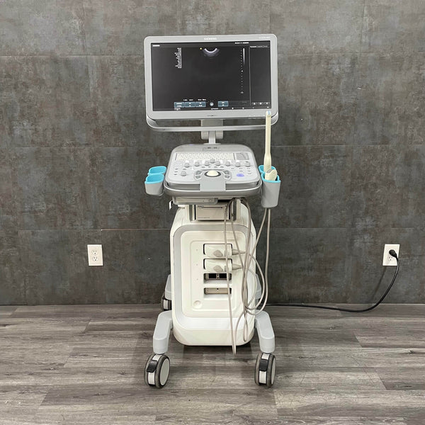
Components of an Ultrasound Machine
- Transducer (Probe): This is the part of the ultrasound machine that the doctor moves over your skin. It’s like the camera lens for the sound waves, sending them out and picking them up when they bounce back.
- Console: Think of this as the brain of the operation. The console has all the controls and settings that let the doctor adjust the ultrasound to get the best picture.
- Display: After the sound waves have their photo session inside your body, the display shows what they've seen. It’s where the images from the sound waves appear, so the doctor can see what’s up.
- Software: This is the smart part that takes the information from the transducer and turns it into an image. Without it, all those echoes would just be noise.
From the transducer that starts the whole process to the software that makes sense of it all, each part of an ultrasound machine has a special role to play in letting doctors see inside you. It's tech that helps keep us healthy, all without a scratch.
Benefits of Using Ultrasound Machines
Ultrasound machines, often known as diagnostic ultrasounds or sonograms, are essential tools in medical imaging that utilize high-frequency sound waves to create detailed images of the internal structures of the body. These images aid in the diagnosis and monitoring of various conditions across different medical fields.
Non-Invasive and Safe
- GE Voluson P8: Like other ultrasound machines, it ensures non-invasive monitoring, which is crucial during pregnancy. Its use in obstetric care demonstrates the safety and effectiveness of ultrasound equipment in tracking fetal development.
- Samsung Medison SonoAce X6: This ultrasound tool provides a comfortable and non-invasive diagnostic experience, which is vital in any medical setting where patient comfort is a top priority.
Real-Time Imaging
- Philips ClearVue 350: Notable for its real-time imaging, this ultrasound machine aids significantly in complex diagnostic procedures, delivering high-resolution visuals instantaneously.
- Siemens Acuson X150: This machine is widely used in echo machines for cardiology, providing real-time imaging that is essential for dynamic studies like echocardiograms.
Diverse Applications
- Sonosite 180 Plus: Its portable ultrasound machine design is perfect for varied medical scenarios, from emergency responses to prenatal care, showcasing the versatility of portable ultrasonography.
- Toshiba Famio 8: Even available as a rental, it fulfills diverse diagnostic needs with its quality imaging, indicative of the robust nature of ultrasound equipment.
Detailed Visualization
- Medison SonoAce X4: This model likely offers 3D/4D imaging, providing detailed visualization crucial in specialties like obstetrics and gynecology — a testament to the advanced capabilities of modern sonography machines.
- Siemens Sonoline Adara: While it may offer more traditional imaging, the detailed views it provides are still invaluable within its application range, maintaining its place as a reliable ultrasound scanner.
Accessibility and Portability
- Sonosite Micro Maxx: Known for its portability, this ultrasound machine can be easily transported to a patient's bedside or to remote locations, enhancing healthcare accessibility.
- Esaote Biosound Portable: This machine's mobility makes it simple to move between rooms or clinics, highlighting the convenience of portable usg machines.
Enhanced Diagnostic Accuracy
- GE Voluson P8: Renowned for its sophisticated imaging capabilities, it plays a critical role in the early detection of fetal anomalies, reflecting the high ultrasonography machine cost associated with advanced ultrasound technology.
- Philips ClearVue 350: With its reputation for crisp, clear images, it contributes to the accuracy of diagnoses in various medical fields, demonstrating the value of quality ultrasound equipment.
Guidance for Procedures
- Siemens Acuson X150: Equipped with needle visualization technology, this machine aids in precision-guided procedures such as biopsies, leveraging the precise nature of ultrasound technology.
- Sonosite 180 Plus: Combines high-quality imaging with portability, offering guidance for bedside procedures and interventions, illustrating the practicality of ultrasound tools in clinical settings.
Each machine listed here exemplifies the key benefits of ultrasound technology, with unique features tailored to specific medical needs. From enhancing the safety of non-invasive diagnostics to providing real-time guidance for procedures, these ultrasound machines contribute to better healthcare outcomes
|
Consideration |
Description |
Machines Reference |
|
Imaging Modes |
Types of imaging (2D, 3D, 4D, Doppler) |
GE Voluson P8 (3D/4D), Siemens Acuson X150 (Doppler) |
|
Portability |
Whether the unit needs to be stationary or portable |
Sonosite 180 Plus, Esaote Biosound (Portable) |
|
Ease of Use |
User interface and software simplicity |
Samsung Medison SonoAce X6 |
|
Price |
Upfront cost and financing options |
Toshiba Famio 8 (Rental), Siemens Acuson X150 (Sale) |
|
Durability |
Build quality and lifespan of the machine |
Sonosite Micro Maxx (known for durability) |
|
Service and Warranty |
Availability and length of warranty; service plans |
Inquire for each model, like Philips ClearVue 350 |
|
Upgradeability |
Ability to update software or add features |
Check for Siemens Sonoline Adara upgrade options |
|
Manufacturer Reputation |
Track record of reliability and customer satisfaction |
GE, Philips, Siemens have strong reputations |
|
Accessory Compatibility |
Availability of compatible probes and accessories |
Medison SonoAce X4 (check for accessories) |
|
Technical Support |
Manufacturer support for technical issues |
Inquire for each model, like GE Voluson P8 |
How to Operate an Ultrasound Machine
In a hospital setting, operating an ultrasound machine efficiently is crucial for routine diagnostics and patient care. These machines, which range from sophisticated echocardiogram machines to versatile portable ultrasound machines, are integral in delivering fast and accurate medical assessments. This section will guide hospital staff through the basics of using these essential pieces of ultrasound equipment, ensuring they can maximize the technology’s capabilities to benefit patient care.
Basic Usage Guidelines
- Setup: Start by ensuring that the ultrasound machine is correctly connected to the power supply. Place it near the patient’s bed, making sure to utilize Medical Chairs or Operating Table for optimal positioning.
- Initialization: Turn on the machine and wait for it to boot up. This is a good time to ensure that all necessary accessories, such as the ultrasound wand or probe, are clean and ready.
- Patient Preparation: Position the patient comfortably, often using items like Tilted Table or Echo Tables depending on the type of examination. Apply a liberal amount of gel to the area to be examined to ensure good contact between the skin and the transducer.
- Image Acquisition: Select the correct settings on the ultrasound console or software based on the type of scan. Carefully move the transducer over the area of interest to capture images.
- Interpretation: View the images on the display. Adjust the settings if needed to get clearer images. Many machines also have Patient Monitor functions to observe vital signs concurrently.
- Documentation: Record the findings and save the images in the hospital’s information system, ensuring compliance with data protection regulations.
Common Procedures
- Pregnancy Check-Ups: Use an obstetrics sonography machine to monitor fetal development and maternal health. This involves regular scans to check the fetus's growth, position, and overall well-being.
- Cardiovascular Assessments: Utilize an echocardiogram machine to assess heart function. These scans are critical for detecting heart diseases and guiding treatment plans.
- Abdominal Scans: Conduct examinations to assess organs like the liver, kidneys, and gallbladder using a general ultrasound scanner. This is essential for diagnosing conditions such as stones, cysts, or other anomalies.
- Vascular Imaging: Apply Doppler ultrasound techniques to evaluate blood flow in arteries and veins. This can help detect blockages or abnormalities in the circulatory system.
- Guided Procedures: For more complex interventions, such as biopsies, use ultrasound-guided techniques to ensure precision. This often involves real-time imaging to guide the needle to the correct location.
Each of these procedures leverages the unique capabilities of various ultrasound tools and machines, from the basic portable usg machine to more advanced echo machines. Proper operation not only improves diagnostic accuracy but also enhances the patient’s safety and comfort during the process.
Maintenance and Care
Ensuring that your ultrasound machine is well-maintained and properly cared for is crucial in a hospital setting, not only to extend the lifespan of the ultrasound equipment but also to guarantee the best diagnostic results and patient safety. This section provides practical guidance on regular maintenance, troubleshooting common issues, and ensuring compliance with safety and data handling standards.
Routine Maintenance
- Daily Cleaning: After each use, clean the ultrasound wand and other surfaces of the ultrasound machine with approved disinfectant wipes to prevent the spread of infection.
- Regular Servicing: Schedule routine checks to ensure that all parts of the ultrasound equipment, including portable ultrasound machines and echo machines, are functioning correctly. This may include calibrations and adjustments by a certified technician. Regular maintenance is also essential for other diagnostic tools such as EKG Machine and Medical Scale to ensure accuracy and reliability.
- Software Updates: Keep the software up-to-date to ensure that your ultrasound machine operates efficiently and incorporates the latest imaging technologies.
Troubleshooting Common Issues
- Image Quality Problems: If the image quality degrades, check for common issues like improper settings or a malfunctioning transducer. Consult the user manual for specific troubleshooting tips related to your model.
- Power Issues: Ensure that all connections are secure, especially in portable usg machines which rely on battery power. Check for any loose cables or failures in the power supply.
- Operational Errors: If the machine stops working or displays error messages, refer to the troubleshooting section of the manual. Resetting the system or contacting technical support may be necessary.
Safety and Compliance
- Regulatory Standards: Always ensure that your ultrasound machine complies with local and international safety standards, such as FDA approval in the U.S. This helps to ensure that the equipment is safe and effective for patient diagnostics.
- Radiation Exposure: It’s important to clarify that ultrasound machines use sound waves, not ionizing radiation. There is no risk of radiation exposure, making ultrasounds a safe diagnostic tool for all patients, including pregnant women.
Integration with Other Medical Systems
- Compatibility: Modern ultrasound machines should integrate seamlessly with hospital systems such as Electronic Health Records (EHRs) and imaging software. This compatibility helps in efficient data management and supports continuity of care.
- Data Handling: Ensure that data from ultrasound scans is securely stored and shared in compliance with health information privacy laws like HIPAA in the U.S. Proper data management not only protects patient privacy but also enhances the usability of diagnostic information across the healthcare system.
Maintaining and caring for your ultrasound equipment is a vital part of hospital operations, ensuring that every sonography machine, from portable sonograms to stationary echocardiogram machines, is ready for use and capable of providing high-quality diagnostics.
When choosing ultrasound machines and cardiology equipment, AngelUS stands out as a top choice for healthcare providers looking for quality, reliability, and innovation. Our comprehensive range of ultrasound equipment, including portable ultrasound machines, echocardiogram machines, and integrated ECGs/EKGs, ensures that medical professionals have access to the best diagnostic tools. AngelUS is dedicated to advancing medical technology and making it accessible to hospitals and clinics worldwide.
Frequently Asked Questions
A: First, ensure that the ultrasound machine is plugged in properly. Check both the power cord and the outlet. If the problem persists, inspect the cord for damage, and try a different outlet to confirm there isn’t an issue with the power supply.
A: Poor image quality on your ultrasound machine can often be resolved by adjusting the settings. Ensure that the ultrasound wand is correctly connected, and check the device settings such as brightness and contrast. Regularly cleaning the wand and using appropriate contact gel can also help improve the clarity of images.
A: Maintain your ultrasound machine by cleaning it with recommended solutions after each use, especially the ultrasound wand and any surfaces. Keep the software updated, and regularly check the connections and cables for signs of wear. It’s also a good idea to schedule professional servicing annually.
A: Regular training for operators of the ultrasound machine can significantly reduce user errors. Ensure that your ultrasound equipment is kept up-to-date with the latest software updates and maintained according to the manufacturer's guidelines. Frequent checks for calibration and alignment can also prevent errors.
A: Slow boot times could be due to outdated software on your ultrasound machine. Try restarting the device to clear temporary files and ensure that all software updates are applied. If slow boot times persist, consider consulting with a technician to check for hardware issues.
A: If the ultrasound probe is not recognized, disconnect it and reconnect it securely. Check for any visible damage to the probe or its connection port. If the issue continues, try using a different probe to determine if the problem is with the probe or the ultrasound scanner itself.
A: When parts of your ultrasound machine, such as the probe or display, start malfunctioning regularly or repairs become frequent, it may be time to consider replacements. If the ultrasonography machine cost of ongoing maintenance outweighs the cost of new equipment, upgrading to a new ultrasound machine for sale might be the best option.