


What is Hysteroscopy?
Hysteroscopy is a minimally invasive endoscopic surgical procedure used for the direct visualization and treatment of intrauterine pathology. Derived from the Greek words "hystero" (uterus) and "skopein" (to look), the term essentially means "looking inside the uterus." The procedure involves the insertion of a hysteroscope a thin, lighted tube with a camera through the cervix into the uterine cavity.
At AngelUS, we are committed to providing the highest quality Hysteroscopes and hysteroscopy instruments to ensure precise diagnosis and effective treatment of intrauterine conditions. Our state-of-the-art technology and ergonomic designs make our products the preferred choice for healthcare professionals worldwide.
Purpose of Hysteroscopy
Diagnostic Hysteroscopy
-
Indications:
- Abnormal uterine bleeding (AUB)
- Infertility and recurrent pregnancy loss
- Suspected intrauterine pathology (e.g., polyps, fibroids)
- Congenital uterine anomalies
- Abnormal uterine bleeding (AUB)
-
Procedure:
- Performed to inspect the endometrial cavity, tubal ostia, and endocervical canal.
- Utilizes a small-caliber (usually <5 mm) hysteroscope, often without the need for anesthesia or with minimal sedation.
- The hysteroscope transmits a high-resolution image of the uterine cavity to an external monitor for detailed examination. AngelUS offer hysteroscopes that are designed to deliver clear, high-definition images, ensuring accurate diagnosis.
- Performed to inspect the endometrial cavity, tubal ostia, and endocervical canal.
Operative (Therapeutic) Hysteroscopy
-
Indications:
- Resection of endometrial polyps or submucosal fibroids (hysteroscopic
- polypectomy, hysteroscopic myomectomy).
- Removal of intrauterine adhesions (Asherman's syndrome)
- Septoplasty for uterine septum
- Endometrial ablation for menorrhagia
-
Procedure:
- Requires a slightly larger hysteroscope (5-10 mm) with working channels for surgical instruments.
- Usually performed under general or regional anesthesia.
- Instruments such as resectoscopes, morcellators, or lasers are used to perform precise surgical interventions.
- Requires a slightly larger hysteroscope (5-10 mm) with working channels for surgical instruments.
AngelUS is your trusted partner for advanced hysteroscopy solutions, ensuring top-quality visualization and treatment for intrauterine conditions.
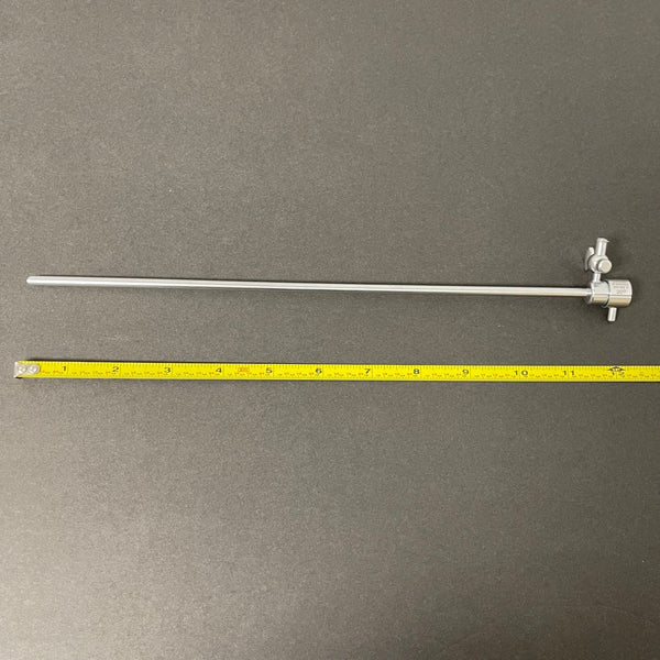
What are Hysteroscope?
A hysteroscope is an advanced endoscopic instrument used for examining and treating the uterine cavity. It comprises several key components, including a high-definition optical system for superior image quality, control mechanisms for precise navigation, an outer sheath to protect internal components, working channels for the insertion of surgical instruments, and a fluid management system to maintain cavity distension and visualization. AngelUS hysteroscopes integrate these features to provide clinicians with reliable and effective tools for both diagnostic and therapeutic hysteroscopic procedures.
Components of a Hysteroscope
-
Optical System: High-definition camera with fiber optics for illumination, like those available in AngelUS hysteroscopes for superior image quality.
-
Control Mechanisms: Ergonomic grips for precise navigation, integrated imaging for real-time video, image, and documentation, all features of AngelUS hysteroscopes.
-
Outer Sheath: Encases and protects internal components, includes channels for saline or CO₂ to expand the uterine cavity.
-
Working Channels: Instrument ports for inserting micro-instruments (scissors, forceps, electrosurgical devices) necessary for therapeutic procedures.
-
Fluid Management System: Ensures cavity expansion and visualization, maintains consistent distension, and minimizes fluid overload.
For a more detailed understanding of each component and its role in hysteroscopy procedures, please refer to our detailed guide on the Components of a Hysteroscope.
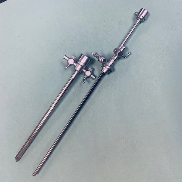
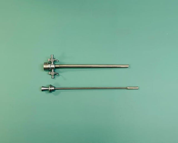
What are the Types of Hysteroscopes?
Diagnostic Hysteroscopes
Diagnostic hysteroscopes are specifically designed for the inspection and evaluation of the uterine cavity. They are typically smaller in diameter than operative hysteroscopes and are primarily used for diagnostic purposes rather than therapeutic interventions.
Features
-
Diameter and Size:
-
Small Caliber: Diagnostic hysteroscopes usually have a diameter ranging from 2.7 mm to 4 mm. The smaller size facilitates easier insertion through the cervical canal with minimal discomfort and often without the need for dilation.
-
Compact Design: Their compact design allows for high maneuverability within the uterine cavity, providing detailed visualization of the endometrium and other intrauterine structures.
-
Small Caliber: Diagnostic hysteroscopes usually have a diameter ranging from 2.7 mm to 4 mm. The smaller size facilitates easier insertion through the cervical canal with minimal discomfort and often without the need for dilation.
-
Optical System:
-
High-Resolution Imaging: Equipped with high-resolution cameras and fiber optic light sources, these hysteroscopes provide clear, magnified Hysteroscope images of the uterine cavity.
-
Variable Angulation: Many diagnostic hysteroscopes offer different viewing angles (e.g., 0°, 12°, 30°) to increase the field of view and allow comprehensive examination of the uterine walls and tubal ostia.
-
High-Resolution Imaging: Equipped with high-resolution cameras and fiber optic light sources, these hysteroscopes provide clear, magnified Hysteroscope images of the uterine cavity.
-
Fluid Management:
-
Distension Media Channels: Diagnostic hysteroscopes have channels for the inflow and outflow of distension media (saline or CO₂), which are essential for expanding the uterine cavity and maintaining a clear view.
-
Distension Media Channels: Diagnostic hysteroscopes have channels for the inflow and outflow of distension media (saline or CO₂), which are essential for expanding the uterine cavity and maintaining a clear view.
-
Ergonomic Design:
- User-Friendly Handpieces: These hysteroscopes often feature ergonomically designed handpieces to facilitate ease of use and precise control during the procedure.
-
Lightweight Construction: Their lightweight construction reduces operator fatigue, allowing for prolonged use during extensive examinations.
-
Integrated Technology:
-
Image and Video Recording: Advanced diagnostic hysteroscopes are integrated with digital systems that enable real-time video streaming, image, and documentation for further analysis and patient records.Including a hysteroscopy diagram can help visualize the procedure and its components.
- Compatibility with Various Monitors: They are compatible with various endoscopic monitors and imaging systems, providing flexibility in clinical settings.
-
Image and Video Recording: Advanced diagnostic hysteroscopes are integrated with digital systems that enable real-time video streaming, image, and documentation for further analysis and patient records.Including a hysteroscopy diagram can help visualize the procedure and its components.
Applications of Hysteroscopy
-
Evaluation of Abnormal Uterine Bleeding (AUB):
- Investigates causes like polyps, fibroids, hyperplasia, or carcinoma.
- Allows direct visualization and targeted biopsies.
- Investigates causes like polyps, fibroids, hyperplasia, or carcinoma.
-
Assessment of Infertility and Recurrent Pregnancy Loss:
- Identifies abnormalities contributing to infertility or pregnancy loss (e.g., septa, adhesions, congenital anomalies).
- Identifies abnormalities contributing to infertility or pregnancy loss (e.g., septa, adhesions, congenital anomalies).
-
Confirmation of Imaging Findings:
- Confirms findings from ultrasound, MRI, or HSG.
- Provides detailed assessment of intrauterine pathology.
- Confirms findings from ultrasound, MRI, or HSG.
-
Investigation of Endometrial Pathology:
- Evaluates endometrial thickening, irregularities, or other abnormalities.
- Includes targeted biopsies to rule out malignancy.
- Evaluates endometrial thickening, irregularities, or other abnormalities.
-
Pre-Operative Planning:
- Aids in planning for gynecological surgeries by mapping the uterine cavity and identifying potential complications.
By understanding the specific features and applications of diagnostic hysteroscopes, clinicians can effectively utilize these instruments to magnify diagnostic accuracy, guide therapeutic decisions, and improve patient outcomes.
Operative Hysteroscopes
Operative hysteroscopes are designed for performing therapeutic interventions within the uterine cavity. They are larger in diameter compared to diagnostic hysteroscopes and come equipped with channels for surgical instruments.
Features
-
Diameter and Size:
-
Larger Caliber: Operative hysteroscopes typically have a diameter ranging from 5 mm to 10 mm. The increased size accommodates the working channels necessary for various surgical instruments.
-
Robust Construction: Designed to withstand the mechanical stresses of surgical procedures, ensuring durability and reliability.
-
Larger Caliber: Operative hysteroscopes typically have a diameter ranging from 5 mm to 10 mm. The increased size accommodates the working channels necessary for various surgical instruments.
-
Optical System:
-
High-Resolution Imaging: Similar to diagnostic hysteroscopes, operative hysteroscopes are equipped with high-definition cameras and fiber optic light sources, providing clear and magnified Hysteroscope images of the surgical field.
-
Wide Field of View: They offer a wider field of view to increase visibility during complex procedures, with various angulation options (0°, 12°, 30°).
-
High-Resolution Imaging: Similar to diagnostic hysteroscopes, operative hysteroscopes are equipped with high-definition cameras and fiber optic light sources, providing clear and magnified Hysteroscope images of the surgical field.
-
Working Channels:
-
Multiple Channels: These hysteroscopes have one or more working channels (typically 1.5 mm to 3 mm in diameter) for the introduction of surgical instruments such as scissors, graspers, biopsy forceps, electrosurgical devices, and laser fibers.
-
Separation of Functions: Channels are separated for distension media and surgical instruments to maintain a clear view and prevent contamination.
-
Multiple Channels: These hysteroscopes have one or more working channels (typically 1.5 mm to 3 mm in diameter) for the introduction of surgical instruments such as scissors, graspers, biopsy forceps, electrosurgical devices, and laser fibers.
-
Fluid Management System:
-
Increased Distension Media Management: Advanced systems to control the flow and pressure of distension media (saline or CO₂) are important for maintaining uterine cavity distension during surgery.
-
Safety Mechanisms: Integrated safety features to monitor and regulate intrauterine pressure, minimizing the risk of fluid overload and other complications.
-
Increased Distension Media Management: Advanced systems to control the flow and pressure of distension media (saline or CO₂) are important for maintaining uterine cavity distension during surgery.
-
Electrosurgical and Laser Integration:
-
Electrosurgical Tools: Operative hysteroscopes are compatible with electrosurgical units that allow for precise cutting, coagulation, and ablation of tissues. These include resectoscopes with monopolar or bipolar energy sources.
-
Laser Compatibility: They can also be used with various laser systems (e.g., CO₂, Nd) for targeted tissue ablation and precise surgical interventions.
-
Electrosurgical Tools: Operative hysteroscopes are compatible with electrosurgical units that allow for precise cutting, coagulation, and ablation of tissues. These include resectoscopes with monopolar or bipolar energy sources.
-
Control and Ergonomics:
-
Advanced Steering Mechanisms: Operative hysteroscopes often feature sophisticated steering and control mechanisms, allowing for precise navigation and manipulation within the uterine cavity.
- Ergonomic Design: User-friendly handgrips and controls to reduce operator fatigue and increase precision during lengthy procedures.
-
Advanced Steering Mechanisms: Operative hysteroscopes often feature sophisticated steering and control mechanisms, allowing for precise navigation and manipulation within the uterine cavity.
Applications of Operative Hysteroscope
-
Resection of Endometrial Polyps and Submucosal Fibroids
-
Procedure: Hysteroscopic Polypectomy and Hysteroscopic myomectomy using electrosurgical loops or mechanical morcellators. Also, Instruments such as the truclear hysteroscope and myosure hysteroscope are often used for removing polyps and fibroids.
-
Indications: Abnormal uterine bleeding, infertility, or recurrent pregnancy loss due to intrauterine growths.
-
Procedure: Hysteroscopic Polypectomy and Hysteroscopic myomectomy using electrosurgical loops or mechanical morcellators. Also, Instruments such as the truclear hysteroscope and myosure hysteroscope are often used for removing polyps and fibroids.
-
Removal of Intrauterine Adhesions (Asherman's Syndrome)
-
Procedure: Adhesiolysis to remove scar tissue and restore normal anatomy.
-
Indications: Amenorrhea, infertility, or recurrent pregnancy loss due to adhesions.
-
Procedure: Adhesiolysis to remove scar tissue and restore normal anatomy.
-
Endometrial Ablation
-
Procedure: Techniques like thermal, electrosurgical, or laser ablation to destroy the endometrial lining.
-
Indications: Menorrhagia unresponsive to medical management in patients not desiring future fertility.
-
Procedure: Techniques like thermal, electrosurgical, or laser ablation to destroy the endometrial lining.
-
Correction of Congenital Uterine Anomalies
-
Procedure: Septoplasty using scissors or electrosurgical instruments to resect a uterine septum.
-
Indications: Infertility or recurrent pregnancy loss due to a confirmed uterine septum.
-
Procedure: Septoplasty using scissors or electrosurgical instruments to resect a uterine septum.
-
Targeted Biopsies and Excision of Pathological Tissue
-
Procedure: Directed biopsies and excision of hyperplastic endometrium or small carcinomas.
-
Indications: Abnormal uterine bleeding, suspected malignancy, or other intrauterine pathology.
-
Procedure: Directed biopsies and excision of hyperplastic endometrium or small carcinomas.
-
Tubal Ostia Cannulation and Fallopian Tube Recanalization
-
Procedure: Cannulation and recanalization of obstructed fallopian tubes.
-
Indications: Proximal tubal obstruction contributing to infertility.
-
Procedure: Cannulation and recanalization of obstructed fallopian tubes.
With AngelUS diagnostic hysteroscopes, experience unparalleled clarity and precision in diagnosing intrauterine conditions.
Flexible vs. Rigid Hysteroscopes
|
Feature/Aspect |
Flexible Hysteroscopes |
Rigid Hysteroscopes |
|
Construction |
Made of flexible materials allowing for bending |
Made of rigid materials, typically stainless steel |
|
Diameter |
Generally smaller diameter (2-5 mm) |
Typically larger diameter (3-10 mm) |
|
Flexibility |
Highly flexible, can guide complex uterine anatomy |
Fixed shape, less flexible |
|
Image Quality |
Slightly lower resolution due to flexible optics |
High-resolution images due to rigid optics |
|
Usage |
Primarily for diagnostic procedures |
Suitable for both diagnostic and operative procedures |
|
Insertion |
Easier insertion, especially in patients with narrow or stenotic cervix |
Requires more dilation for insertion |
|
Patient Comfort |
Generally more comfortable for patients |
May cause more discomfort during insertion |
|
Control Mechanisms |
Limited control over tip direction |
Precise control over tip and instrument manipulation |
|
Durability |
Less durable, more prone to damage |
Highly durable, less prone to damage |
|
Cost |
Generally more expensive due to advanced materials and technology |
Generally less expensive |
|
Application Examples |
Ideal for navigating complex or tortuous anatomy, diagnosing intrauterine conditions |
Used for a wide range of diagnostic and therapeutic procedures, including polypectomy, myomectomy, and adhesiolysis |
|
Steering Mechanisms |
Built-in steering mechanisms for navigating |
No built-in steering; relies on rigid manipulation |
|
Learning Curve |
May require more skill to master due to flexibility |
Easier to learn for beginners due to fixed structure |
Flexible hysteroscopy involves the use of a flexible hysteroscope for better navigation through the uterine cavity as Flexible hysteroscopes are made of flexible materials allowing for bending, while rigid hysteroscopes are made of stainless steel.
Applications of Hysteroscopy
Diagnostic Applications
Hysteroscopy is a valuable diagnostic tool in gynecology. It allows direct visualization of the uterine cavity, providing detailed information that is essential for diagnosing various uterine conditions. Here are the main diagnostic applications:
Identifying Uterine Abnormalities
-
Abnormal Uterine Bleeding (AUB):
-
Description: AUB refers to any deviation from the normal menstrual cycle, including heavy bleeding, prolonged bleeding, or irregular cycles.
-
Role of Hysteroscopy: Hysteroscopy enables direct visualization of the endometrial cavity to identify the source of abnormal bleeding. Conditions such as endometrial polyps, submucosal fibroids, endometrial hyperplasia, and carcinoma can be detected and assessed.
-
Process: During the procedure, the hysteroscope is inserted through the cervix into the uterine cavity. The uterine cavity is then distended using saline or CO₂, allowing the physician to inspect the endometrial lining for abnormalities.
-
Description: AUB refers to any deviation from the normal menstrual cycle, including heavy bleeding, prolonged bleeding, or irregular cycles.
-
Endometrial Polyps:
-
Description: Polyps are benign growths of the endometrial tissue that can cause irregular bleeding and infertility.
-
Role of Hysteroscopy: Hysteroscopy allows for the direct visualization and precise localization of polyps. This is important for planning their removal.Hysteroscopic laparoscopy combines hysteroscopy and laparoscopy for comprehensive treatment.
-
Process: The hysteroscope helps identify the number, size, and location of polyps, which can be targeted for subsequent removal using operative hysteroscopy.
-
Description: Polyps are benign growths of the endometrial tissue that can cause irregular bleeding and infertility.
-
Submucosal Fibroids:
-
Description: These are benign tumors of the smooth muscle within the uterine wall that protrude into the uterine cavity, causing heavy menstrual bleeding and fertility issues.
-
Role of Hysteroscopy: Hysteroscopy provides a clear view of the fibroids' size, location, and impact on the uterine cavity.
-
Process: Fibroids are visualized, and their relationship with the endometrial lining and uterine cavity is assessed to guide surgical planning.
-
Description: These are benign tumors of the smooth muscle within the uterine wall that protrude into the uterine cavity, causing heavy menstrual bleeding and fertility issues.
-
Intrauterine Adhesions (Asherman’s Syndrome):
-
Description: Adhesions are bands of scar tissue within the uterine cavity, often resulting from previous surgeries or infections, leading to menstrual irregularities and infertility.
-
Role of Hysteroscopy: Hysteroscopy is the standard for diagnosing intrauterine adhesions, as it allows direct visualization and assessment of the extent of adhesions.
-
Process: The adhesions are identified and evaluated for their impact on the uterine cavity's shape and function.
-
Description: Adhesions are bands of scar tissue within the uterine cavity, often resulting from previous surgeries or infections, leading to menstrual irregularities and infertility.
-
Congenital Uterine Anomalies:
-
Description: These are structural abnormalities of the uterus present from birth, such as a septate uterus, bicornuate uterus, or arcuate uterus.
-
Role of Hysteroscopy: Hysteroscopy allows detailed examination of the uterine cavity to diagnose congenital anomalies accurately.
-
Process: The shape and structure of the uterine cavity are inspected to identify anomalies that may affect fertility and pregnancy outcomes.
-
Description: These are structural abnormalities of the uterus present from birth, such as a septate uterus, bicornuate uterus, or arcuate uterus.
Biopsy Procedures
-
Endometrial Biopsy:
-
Description: An endometrial biopsy involves taking a small sample of the endometrial tissue for histological examination.
-
Role of Hysteroscopy: Hysteroscopy-guided biopsy provides targeted sampling of suspicious areas, increasing the diagnostic accuracy compared to blind biopsy techniques.
-
Process: Under direct visualization, the hysteroscope guides a biopsy instrument to the specific area of interest, ensuring precise tissue sampling.
-
Description: An endometrial biopsy involves taking a small sample of the endometrial tissue for histological examination.
-
Evaluation of Endometrial Hyperplasia:
-
Description: Endometrial hyperplasia is a condition characterized by the thickening of the endometrial lining, which can be a precursor to endometrial cancer.
-
Role of Hysteroscopy: Hysteroscopy allows the direct visualization of the hyperplastic areas and targeted biopsy to confirm the diagnosis and rule out malignancy.
-
Process: The hysteroscope is used to identify areas of thickened endometrium, and biopsy instruments are introduced to obtain tissue samples for histopathological analysis.
-
Description: Endometrial hyperplasia is a condition characterized by the thickening of the endometrial lining, which can be a precursor to endometrial cancer.
-
Suspected Endometrial Carcinoma:
-
Description: Endometrial carcinoma is a malignant tumor of the endometrium, often presenting with abnormal uterine bleeding.
-
Role of Hysteroscopy: Hysteroscopy provides detailed visualization and the ability to perform targeted biopsies of suspicious lesions, improving diagnostic accuracy and aiding in staging the disease.
- Process: Suspicious lesions are identified using the hysteroscope, and biopsies are taken from these areas to confirm malignancy and assess the extent of the disease.
-
Description: Endometrial carcinoma is a malignant tumor of the endometrium, often presenting with abnormal uterine bleeding.
Operative Applications
Removal of Polyps and Fibroids
-
Endometrial Polyps:
-
Description: Endometrial polyps are benign growths of the endometrial tissue that protrude into the uterine cavity. They can cause abnormal uterine bleeding, infertility, and recurrent pregnancy loss.
-
Procedure:
-
Polypectomy: The removal of endometrial polyps is known as polypectomy. This is typically performed using a hysteroscope equipped with mechanical or electrosurgical instruments.
-
Steps:
-
Visualization: The hysteroscope is inserted through the cervix, and the uterine cavity is distended using saline or CO₂.
-
Localization: Polyps are identified and their size and location are assessed.
-
Resection: Instruments such as scissors, forceps, or electrosurgical loops are introduced through the working channels to excise the polyps.
-
Removal: The excised polyps are removed from the uterine cavity for histopathological examination.
-
Visualization: The hysteroscope is inserted through the cervix, and the uterine cavity is distended using saline or CO₂.
-
Polypectomy: The removal of endometrial polyps is known as polypectomy. This is typically performed using a hysteroscope equipped with mechanical or electrosurgical instruments.
-
Description: Endometrial polyps are benign growths of the endometrial tissue that protrude into the uterine cavity. They can cause abnormal uterine bleeding, infertility, and recurrent pregnancy loss.
-
Submucosal Fibroids:
-
Description: Submucosal fibroids are benign smooth muscle tumors located within the uterine wall, protruding into the uterine cavity. They can cause heavy menstrual bleeding, pain, and infertility.
-
Procedure:
-
Myomectomy: The removal of submucosal fibroids is called myomectomy. This can be performed hysteroscopically using electrosurgical or mechanical morcellation techniques.
-
Steps:
-
Visualization: The hysteroscope is inserted and the uterine cavity is distended.
-
Localization: Fibroids are visualized and their size and position are determined.
-
Resection: Electrosurgical loops or morcellators are used to dissect the fibroids from the uterine wall.
-
Removal: The fibroid fragments are removed from the cavity, ensuring complete resection to prevent recurrence.
-
Visualization: The hysteroscope is inserted and the uterine cavity is distended.
-
Myomectomy: The removal of submucosal fibroids is called myomectomy. This can be performed hysteroscopically using electrosurgical or mechanical morcellation techniques.
-
Description: Submucosal fibroids are benign smooth muscle tumors located within the uterine wall, protruding into the uterine cavity. They can cause heavy menstrual bleeding, pain, and infertility.
Treatment of Uterine Septa
Uterine Septum
-
Description: A uterine septum is a congenital malformation where a fibrous or muscular partition divides the uterine cavity partially or completely. It can lead to infertility, recurrent pregnancy loss, and obstetric complications.
-
Procedure:
-
Septoplasty: The surgical correction of a uterine septum is called septoplasty. This procedure is performed hysteroscopically using cutting instruments or electrosurgery.
-
Steps:
-
Visualization: The hysteroscope is inserted and the septum is visualized.
-
Localization: The extent and type of the septum (fibrous or muscular) are assessed.
-
Resection: Scissors, electrosurgical devices, or laser fibers are used to incise and remove the septum.
- Restoration: The uterine cavity is reshaped to a more normal configuration, improving reproductive outcomes.
-
Visualization: The hysteroscope is inserted and the septum is visualized.
-
Septoplasty: The surgical correction of a uterine septum is called septoplasty. This procedure is performed hysteroscopically using cutting instruments or electrosurgery.
Endometrial Ablation
-
Description: Endometrial ablation is a procedure aimed at destroying the endometrial lining to treat heavy menstrual bleeding (menorrhagia). It is considered when medical management fails, and the patient does not wish to preserve fertility.
-
Procedure:
-
Techniques: Several techniques are used for endometrial ablation, including thermal, radiofrequency, microwave, cryoablation, and electrosurgical methods.
-
Steps:
-
Visualization: The hysteroscope is inserted to inspect the endometrial cavity.
-
Preparation: The uterine cavity is distended, and the appropriate ablation device is introduced.
-
Ablation: The selected method is used to uniformly destroy the endometrial lining:
-
Thermal Balloon: A balloon filled with heated fluid is inserted and inflated to apply thermal energy to the endometrium.
-
Radiofrequency: A device delivering radiofrequency energy is used to ablate the endometrial tissue.
-
Microwave: Microwave energy is applied to the endometrium via a specialized probe.
-
Cryoablation: A cryoprobe is used to freeze the endometrial tissue.
-
Electrosurgical: Rollerball or loop electrodes are used to coagulate and destroy the endometrium.
-
Thermal Balloon: A balloon filled with heated fluid is inserted and inflated to apply thermal energy to the endometrium.
-
Completion: The uterine cavity is inspected to ensure complete ablation, and the hysteroscope is removed.
-
Visualization: The hysteroscope is inserted to inspect the endometrial cavity.
-
Techniques: Several techniques are used for endometrial ablation, including thermal, radiofrequency, microwave, cryoablation, and electrosurgical methods.
What are the Advantages of Using a Hysteroscope?
Minimally Invasive Nature
Hysteroscopic surgery provides direct visualization and treatment of intrauterine pathology.The hysteroscope is inserted through the natural opening of the cervix, eliminating the need for open surgery.
-
Less Pain and Discomfort: Patients experience less postoperative pain compared to traditional surgical methods.
- No Visible Scarring: Since there are no large incisions, patients do not have visible scars, which is aesthetically favorable and reduces recovery-related concerns.
Precision and Accuracy
Hysteroscopy instruments provide high-resolution, real-time images of the uterine cavity.
-
Accurate Diagnoses: Direct visualization helps in accurately identifying issues such as polyps, fibroids, and adhesions.
- Targeted Treatment: Surgeons can precisely target abnormal tissues, minimizing damage to surrounding healthy tissues and ensuring effective treatment.
Faster Recovery Times
Due to the minimally invasive nature of hysteroscopy, patients generally recover much faster than they would from traditional open surgery.
-
Shorter Hospital Stays: Many hysteroscopic procedures are performed on an outpatient basis, allowing patients to go home the same day.
- Quick Return to Daily Activities: Patients can resume their normal activities much sooner, reducing downtime and the impact on their daily lives.
Reduced Risk of Complications
The minimally invasive approach of hysteroscopy significantly reduces the risk of complications associated with open surgery, such as infections, bleeding, and long-term adhesions.
-
Lower Infection Rates: The reduced need for large incisions decreases the risk of postoperative infections.
- Minimal Blood Loss: Hysteroscopic procedures involve minimal blood loss, which is safer for the patient and reduces the need for transfusions.
Increased Visualization
The high-definition cameras and fiber optic light sources in hysteroscopes provide excellent visualization of the uterine cavity, enabling detailed examination and precise surgical interventions.
-
Comprehensive Examination: Surgeons can thoroughly inspect the uterine cavity, ensuring no abnormalities are missed.
- Improved Surgical Outcomes: Increased visualization allows for meticulous surgical work, improving the overall success rate of procedures.
By understanding these advantages, users can appreciate the significant benefits of hysteroscopy, making it a preferred choice for both diagnostic and operative procedures within the uterine cavity.
Procedure Overview
Hysteroscopy is a minimally invasive procedure that allows direct visualization and treatment of the uterine cavity. This technique is used for both diagnostic and operative purposes, making it a versatile tool in gynecological practice. During a hysteroscopy, a hysteroscope is inserted through the cervix into the uterus, providing real-time images that guide the diagnosis and treatment of various intrauterine conditions.
For more detailed information on the procedure of hysteroscopy, including step-by-step guidelines and best practices, you can refer to our Detailed Guide on the Procedure of Hysteroscopy.
Safety and Precautions
Common Risks and Complications
While hysteroscopy is generally considered a safe procedure, like any medical intervention, it carries certain risks and potential complications. Understanding these risks and the precautions taken to mitigate them is important for ensuring patient safety.
Infection
-
Description: Infection can occur when bacteria enter the uterine cavity during the hysteroscopy procedure. Though rare, infections can range from mild to severe.
-
Risks and Complications:
-
Endometritis: Infection of the endometrial lining, leading to pelvic pain, fever, and abnormal discharge.
-
Pelvic Inflammatory Disease (PID): An infection that can spread to the reproductive organs, potentially causing chronic pain and infertility.
-
Endometritis: Infection of the endometrial lining, leading to pelvic pain, fever, and abnormal discharge.
-
Precautions:
-
Sterile Technique: Maintaining a sterile environment and using sterile instruments to minimize the risk of introducing bacteria.Hysteroscopy machine and hysteroscopy equipment must be sterile to minimize the risk of infection.
-
Prophylactic Antibiotics: Administering antibiotics before the procedure in high-risk patients to prevent infection.
-
Monitoring: Close postoperative monitoring for signs of infection, such as fever, pain, or unusual discharge, allowing for prompt treatment if an infection develops.
-
Sterile Technique: Maintaining a sterile environment and using sterile instruments to minimize the risk of introducing bacteria.Hysteroscopy machine and hysteroscopy equipment must be sterile to minimize the risk of infection.
Bleeding
-
Description: Bleeding can occur during or after hysteroscopy due to trauma to the endometrial lining or blood vessels within the uterine cavity.
-
Risks and Complications:
-
Minor Bleeding: Small amounts of bleeding are common and usually self-limited.
-
Hemorrhage: Severe bleeding, though rare, can occur and may require additional intervention.
-
Minor Bleeding: Small amounts of bleeding are common and usually self-limited.
-
Precautions:
-
Careful Technique: Using gentle and precise surgical techniques to minimize trauma to the endometrial lining and blood vessels.Proper handling of hysteroscopy instruments is important for patient safety.
-
Electrosurgical Devices: Using electrosurgical instruments to coagulate (seal) blood vessels during the procedure, reducing the risk of significant bleeding.
- Postoperative Monitoring: Observing the patient for signs of excessive bleeding and intervening promptly if it occurs.
-
Careful Technique: Using gentle and precise surgical techniques to minimize trauma to the endometrial lining and blood vessels.Proper handling of hysteroscopy instruments is important for patient safety.
Perforation
-
Description: Uterine perforation is a rare but serious complication where the hysteroscope or surgical instrument punctures the uterine wall, potentially causing injury to adjacent organs.
-
Risks and Complications:
-
Immediate Injury: Damage to the uterus, bladder, bowel, or blood vessels, which may require additional surgical repair.
-
Delayed Complications: Risk of infection, bleeding, or formation of adhesions.
-
Immediate Injury: Damage to the uterus, bladder, bowel, or blood vessels, which may require additional surgical repair.
-
Precautions:
-
Preoperative Assessment: Evaluating the patient's anatomy and medical history to identify any factors that may increase the risk of perforation.
-
Experienced Operators: Ensuring that the procedure is performed by skilled and experienced surgeons who are adept at hysteroscopic techniques.
-
Gentle Manipulation: Using controlled and gentle manipulation of the hysteroscope and instruments to avoid excessive force that could lead to perforation.
- Immediate Response: Being prepared to recognize and manage a perforation immediately if it occurs, including possible laparoscopy or laparotomy to repair the injury.
-
Preoperative Assessment: Evaluating the patient's anatomy and medical history to identify any factors that may increase the risk of perforation.
While hysteroscopy is a minimally invasive and generally safe procedure, awareness and management of potential risks and complications are essential. Key safety measures include maintaining sterile techniques, careful surgical manipulation, and close postoperative monitoring. By understanding these risks and the precautions taken to mitigate them, healthcare providers can increase patient safety and outcomes.
Conditions Where Hysteroscopy is Not Advised
Hysteroscopy is generally a safe procedure, but there are certain conditions where it is contraindicated due to the increased risk of complications or the potential for exacerbating existing conditions. These include:
-
Active Pelvic Infection:
-
Description: Patients with active pelvic inflammatory disease (PID) or other pelvic infections are at higher risk of spreading the infection during hysteroscopy.
-
Reason: Introducing instruments into the uterine cavity can exacerbate the infection and lead to more severe pelvic or systemic infections.
-
Description: Patients with active pelvic inflammatory disease (PID) or other pelvic infections are at higher risk of spreading the infection during hysteroscopy.
-
Known or Suspected Pregnancy:
-
Description: Performing hysteroscopy during pregnancy can disrupt the pregnancy and lead to miscarriage.
-
Reason: The procedure involves distending the uterine cavity and manipulating the endometrial lining, which can adversely affect the developing fetus and pregnancy.
-
Description: Performing hysteroscopy during pregnancy can disrupt the pregnancy and lead to miscarriage.
-
Recent Uterine Surgery:
-
Description: Patients who have recently undergone uterine surgery or procedures (such as cesarean section, myomectomy, or D&C) may have healing tissues that are more susceptible to damage.
-
Reason: The risk of uterine perforation or other complications is higher in the early postoperative period when the tissues are still healing.
-
Description: Patients who have recently undergone uterine surgery or procedures (such as cesarean section, myomectomy, or D&C) may have healing tissues that are more susceptible to damage.
-
Severe Cervical Stenosis:
-
Description: Patients with severe cervical stenosis (narrowing of the cervix) may pose technical challenges for the hysteroscope insertion.
-
Reason: Attempting to force the hysteroscope through a severely stenotic cervix can cause trauma, perforation, or excessive bleeding.
-
Description: Patients with severe cervical stenosis (narrowing of the cervix) may pose technical challenges for the hysteroscope insertion.
-
Known Endometrial or Cervical Malignancy:
-
Description: While hysteroscopy can be used for biopsy and diagnosis, it is generally not performed for definitive treatment of known malignancies due to the risk of spreading cancer cells.
- Reason: Manipulation of malignant tissues can lead to dissemination of cancer cells within the uterine cavity or beyond.
-
Description: While hysteroscopy can be used for biopsy and diagnosis, it is generally not performed for definitive treatment of known malignancies due to the risk of spreading cancer cells.
Discover how AngelUS can increase your practice with our cutting-edge hysteroscopy solutions. Reach out to us for more information, or to place an order.
Frequently Asked Questions
What is the recovery time after a hysteroscopic procedure?
Recovery time is typically short, with most patients resuming normal activities within a day or two. However, this can vary depending on the specific procedure and individual patient factors.
Are there any specific preparations required before undergoing a hysteroscopic procedure?
Patients may be advised to avoid eating or drinking for several hours before the procedure. Additionally, a pre-procedure evaluation including a pelvic exam and possibly imaging studies is usually conducted.
Can hysteroscopy be performed in an office setting, or does it require a hospital visit?
Diagnostic hysteroscopy can often be performed in an office setting without the need for general anesthesia. Operative hysteroscopy, depending on the complexity, may require a hospital or outpatient surgical center.
Is hysteroscopy painful?
Diagnostic hysteroscopy is generally well-tolerated and can be performed with minimal or no anesthesia. Operative hysteroscopy may involve more discomfort and typically requires sedation or anesthesia.
What are the common symptoms that might necessitate a hysteroscopy?
Symptoms include abnormal uterine bleeding, unexplained infertility, recurrent pregnancy loss, pelvic pain, and postmenopausal bleeding.
What is hysteroscopic surgery?
It is a minimally invasive procedure used for diagnosing and treating intrauterine conditions.